how do they x ray babies hips
When the X-rays pass through the body they create an image like a shadow. If it persists they may put on a spica cast.

Developmental Dysplasia Of The Hip Ddh Diagnostic Imaging Developmental Dysplasia Of The Hip Diagnostic Imaging Case Study
About one in eight scans ordered for kids is a CT scan.

. How Do They Xray Babies Head. The frog leg lateral view is a special radiograph of the pelvis to evaluate the hip. Risk factors include 14.
Dense body parts such as bones block the passage of the X-ray beam through the body. Totaleclipse 07092007 1731. X-rays are forms of radiant energy like light or radio waves.
You would have to x-ray your arm or leg more than 5000. 37 years average 5 years. The hip ultrasound will show the healthcare provider the position and shape of the hip joint.
Whilst the parent puts on a lead gown it is the radiographers responsibility to ensure the baby does not roll off the x-ray table. It occurs more commonly in boys typically between 5 and 8 years of age but may range from the ages 3-12. A pelvic X-ray can help your doctor detect various conditions such as.
Your child may be required to hold his or her breath or remain still. Perthes disease also known as Legg-Calvé-Perthes disease is an idiopathic avascular necrosis of the proximal femoral epiphysis. Pregnancy is a time to take good care of yourself and your unborn child.
Appointments and Referrals. X-rays have more energy than rays. X-ray examinations are usually quick and simple.
They give your healthcare provider information about structures inside the body. Its a cast that goes around both hips and down the leg to keep the hips aligned. An X-ray is a safe and painless test that uses a small amount of radiation to make an image of bones organs and other parts of the body.
X-Rays Pregnancy and You. The age groups were based on exposures suitable for tissue thickness in the direction of the X-ray beam of a patient of averagestandard size in that age group for each projection. An X-ray is a picture which is taken using a form of radiation that is able to pass through the body to create a digital X-ray image.
Arthritis that affects your hip. X-rays are a kind of imaging test. The black-and-white images show the internal structures of the hip including the ball-shaped top of the thighbone femoral head and its socket acetabulum in the pelvic bone.
The scan can be done on babies up to about 6 months of age. An ultrasound machine sends sound waves into the hip area and images are recorded on a computer. Inflammation where your sacrum joins the ilium which is called sacroiliitis.
If a child is older than 3 months or 13 weeks then an alpha angle of 50-59 degrees is. A type IIa- hip is at risk to develop dysplasia. 40-240 millirad for an x-ray of the pelvis.
You will go in the room with him he will need to be stripped from the waist down they will take x-rays of him flat on his back legs dead straight and together you wil be able to hold him in this position then an x-ray of his still on his back with his knees bent facing outwards and the soles of his. Many things are especially important during pregnancy such as eating right cutting out. 200-245 millirad for an x-ray of the abdomen.
So an alpha angle of 56 degrees at the age of 7 weeks is called type IIa while at the age of 10 weeks it is called a type IIa-. The American Academy of Pediatrics does not recommend routine ultrasounds for every infant. It can occur bilaterally but it is usually asymmetric.
Around 6 months of age enough bone is present in an infant hip to make an X-ray more accurate than ultrasound. 812 years average 10 years. Once the radiographer a person specially trained in taking X-ray images has positioned the part of your childs body to be examined and lined up the X-ray machine the X-ray examination takes less than a second to perform.
Because they spin around the body taking multiple images CT scans can deliver radiation doses that are up to 200 times higher than an. Different parts of the body contain different tissues which vary in how much X-ray radiation they absorb depending on how dense they are. If she does have it they may try to brace it first.
It is put on by an orthopedic surgeon while using x-ray to make sure the hip is aligned correctly. 51-370 millirad for x-rays of the hip and femur thighbone It is very rare for a single diagnostic x-ray to exceed even 5 rad. The X-ray image is black and white.
But for babies with an abnormal physical exam or major risk factors for developmental dysplasia of the hip or DDH family history Breech position etc the AAP supports referral for. These look white on the X-ray image. These tests expose children to low doses of radiation.
1317 years average 15 years. Is extremely beneficial for pelvis imaging as young children will often begin to cry the moment they are placed supine. The reported incidence of developmental dysplasia of the hip varies between 15 and 20 per 1000 births 1 with the majority 60-80 of abnormal hips resolving spontaneously within 2-8 weeks 1 so-called immature hip.

Pin On Kallie Mae Pup

This Is An X Ray Of A 2 Week Old Puppy Look At How Far The Bones Have To Grow Before They Become A Proper Bony Joint This Is Why Puppies

X Ray Image Of Child Swallowed The Coins For A Medical Diagnosis Medicine Pictures Children Images X Ray Images

My Hips Pre Pao Rpao January 2011 Rpao January 2011 Screws From My Rpao X Ray Ehlers Danlos Syndrome Surgery Recovery

Pin On X Rays

Pin On X Rays

Pin On Nursing 1st Semester

Pin On Radiology Aka X Rays

Pin On X Ray Imaging

Pin On Hip Dysplasia Pao

Anatomy And Physiology Anatomy Sacroiliac

Pin On Adult Hip Dysplasia Awareness
Pin By Meg Carter On Ortho Hip Dysplasia X Ray Orthopedics

The Importance Of Good Positioning On Canine Hip X Rays Hips X Ray Canine

Pin On درمانی
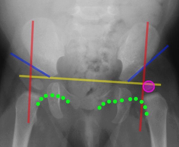
Lines Of The Hip Pediatrics Pediatrics Pediatric Nurse Practitioner Pediatric Radiology

Anatomy Pathology Medicine Nursing Radiography Radiologictechnologist Radiology Radiologystudent Instagram Medical Anatomy Radiology Student Radiology

Hip Dysplasia When You Re Too Young For A Hip Replacement Periacetabular Osteotomy Pao Hip Replacement Recovery Hip Replacement Bursitis Hip

Pin On Pavlik Harness